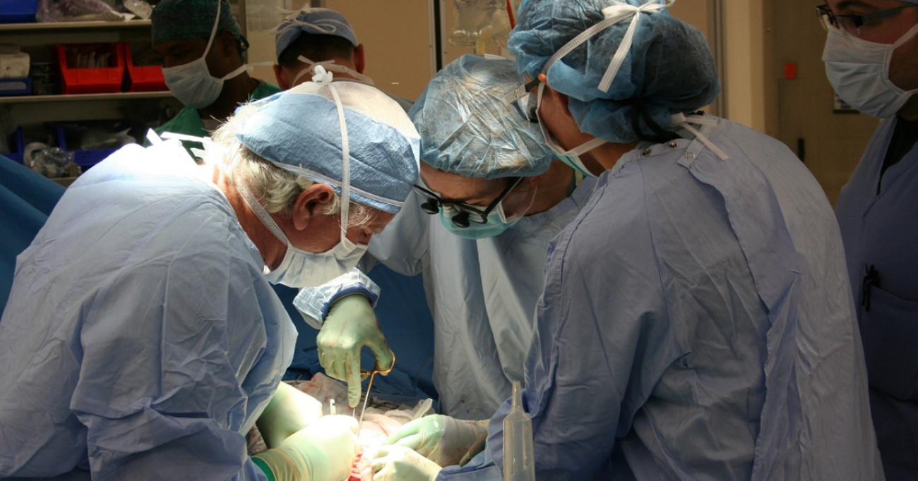
Janelle Letzen, una ricercatrice in psicologia clinica presso la Johns Hopkins University di Baltimora, negli Stati Uniti, a gennaio 2018 ha deciso di unire la scienza alla sua passione: il sushi.
Lo ha fatto usando il suo account Instagram “the_sushi_scientist” , nel quale, tra tonno, avocado, riso, e alghe, spiega visivamente argomenti che vanno dalla neuroscienza alla geologia.
L’amore della dottoressa Letzen per il sushi è iniziato nel 2017 e subito capì che avrebbe potuto combinare insieme concetti scientifici con composizioni culinarie.
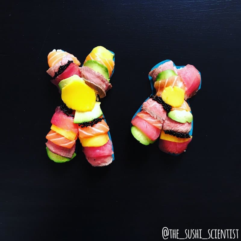
Uno degli obiettivi dello studio della dottoressa Letzen è quello usare il sushi per spiegare le nuove politiche del National Institutes of Health sul sesso come variabile biologica nella ricerca
L’intuizione di Letzen si inserisce in una pratica più ampia, chiamata “Scienstagramm”, che consiste nell’utilizzare il famoso social network per informare visivamente il pubblico su argomenti che altrimenti sarebbero di difficile spiegazione.
Grazie a questa pratica, gli scienziati rendono la loro materia accessibile e comprensibile.
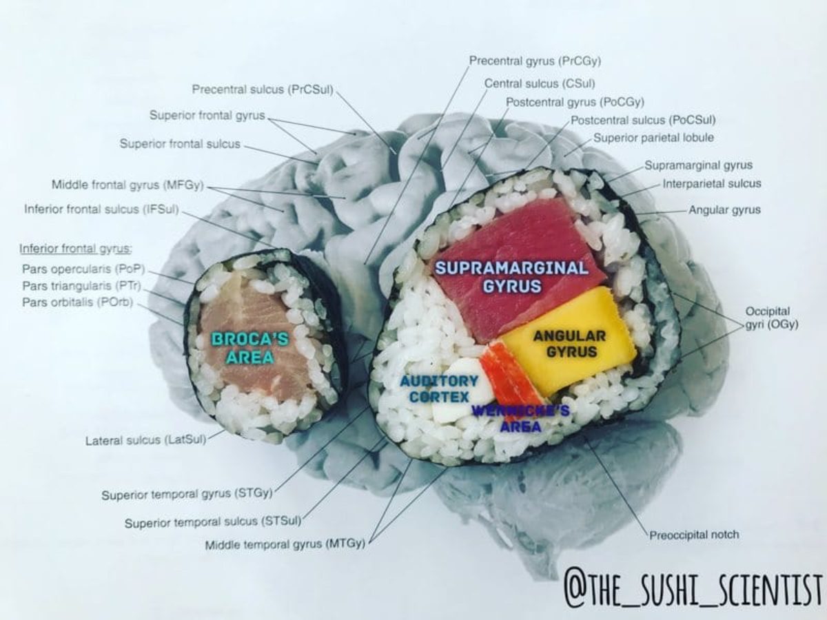
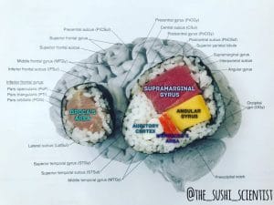
Le sezioni del cervello che controllano il linguaggio, raffigurate usando pesce e riso
Letzen è convinta che per ora i suoi followers siano per lo più professionisti della medicina e studenti interessati alla biopsicologia e alle neuroscienze.
“Ma sto anche cercando di attirare l’attenzione di altri studenti, rendendo la scienza più tangibile”, ha raccontato al portale AtlasObscura .
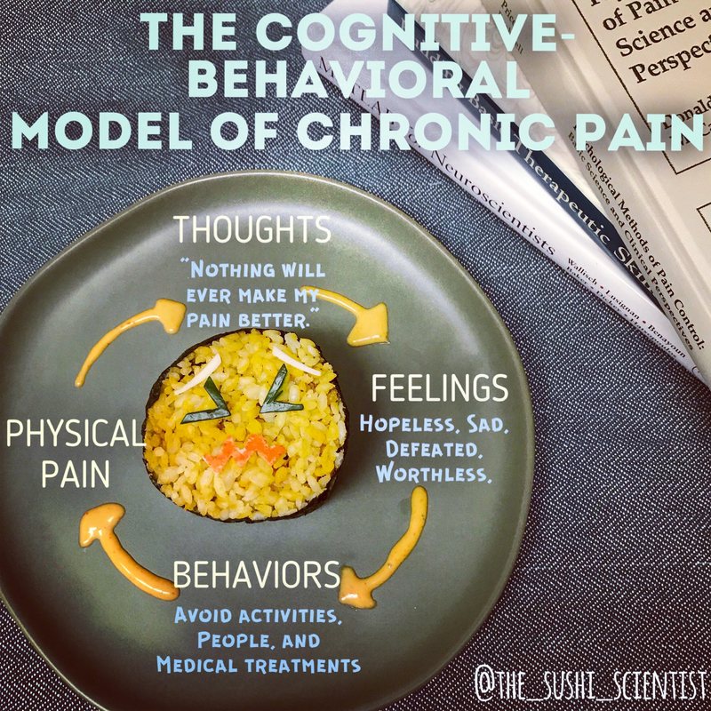
Il piatto spiega la connessione tra i meccanismi neurali e il dolore cronico
Uno dei migliori esempi del lavoro della dottoressa è stato un video di un cervello fatto di sashimi che ha subito una commozione cerebrale.
Le creazioni di Letzen generano anche dibattito.
In un post che aveva come tema gli effetti delle sostanze allucinogene sul cervello umano, creato grazie a rotoli di sushi dai colori vivaci disposti su un piatto, uno studente ha chiesto a Letzen di spiegare l’efficacia di sostanze psichedeliche come l’MDMA per fini terapeutici.
UPDATED: “Hallucinogens” are drugs that cause profound changes in a person’s perception of reality (i.e., hallucinations). They make people hear sounds, see images, and/or feel sensations that are not real. The seared tofu compound above demonstrates phencyclidine, commonly known as PCP in the US. It was originally developed as an anesthetic, but discontinued in the 1960s. It is not commonly used in the US anymore. * Other hallucinogens include LSD, peyote, and psilocybin (i.e., magic mushrooms). Although most of these drugs still remain illegal in the US, ongoing clinical trials have demonstrated efficacy of various hallucinogens in the treatment of addiction, post traumatic stress disorder, depression, and other mental health conditions. * For example, Carhart-Harris et al. (2016, The Lancet) demonstrated that high doses of psilocybin were moderately effective in reducing depression scores even after 3 months of administration in 12 patients with unipolar treatment-resistant depression. Psilocybin acts as a serotonin receptor agonist (see post from 1/18/18 for a demonstration). Some participants reported temporary side effects, like increased anxiety at the onset of the drug’s effects, transient confusion, disordered thinking, mild nausea, and headache. The authors determined that the efficacy on depression symptoms highly outweighed these temporary effects. * When taken in controlled settings and doses, hallucinogens might prove efficacious in treating a wide variety of mental health conditions. Luckily, more research is ongoing in this exciting field. ************* #science #sciencedaily #sciart #sushiart #studygram #chemistry #pharmacy #medicalstudents #md #psychology #phd #neuroscience #nursing #rn
Un post condiviso da The Sushi Scientist (@the_sushi_scientist) in data: Apr 10, 2018 at 4:10 PDT
A quel punto, la dottoressa ha contattato un esperto sull’argomento e ha aggiornato il testo del post “per migliorarlo”.
Ma la domanda che Letzen riceve più spesso è una: “Cosa ne fa del sushi dopo averlo fotografato?”.
La risposta è semplice “Me lo mangio!”, dice Letzen.
Ecco alcuni esempi del lavoro della dottoressa Letzen tratti dal suo profilo Instagram:
Serving up some thalamus nigiri for this #tastytuesday! The thalamus is a structure positioned right above the brainstem. Its main function is to act as a “sensory relay station,” meaning it takes incoming sensory information and appropriately sends it to the cerebral cortex for complex processing. Read below to learn more about how the thalamus works, and comment or DM with topic requests! * * * 1. Let’s think about the cerebral cortex as executive board members for the company, Brain Co. This company’s goal is to take things that people see, hear, smell, and touch to create personalized experiences. These experiences include things like emotions, thoughts, and physical movements. The board members want to decide how these personalized experiences turn out, but they need a way of managing the overwhelming amount of sensory information presented to their company. . * 2. Brain Co. has several stores that are each specialized in collecting different types of sensory information. To run efficiently, they need a regional manager that will assess the stores’ work to decide which cerebral cortex board member would be most interested in this type of information for experience personalization. The thalamus acts as Brain Co.’s regional manager. * 3. In this role, the thalamus has a handy organizational scheme to compartmentalize overwhelming amounts of information. The thalamus organizes all of these different sensory inputs using “nuclei,” or bundles of specialized neurons that differ slightly among each nucleus. . * 4. There might be ~50 specialized thalamic nuclei, but the most popular ones are served up in this nigiri. Further, each nucleus deals with potentially more than one type of sensory information, so the most well-established are listed above. . * 5. If you swipe through the next two pictures, you’ll see where the thalamus is positioned in the brain (Fig. 2), and Brain Co.’s organizational structure for different types of information laid out in “cortico-basal loops” (Fig. 3). *************************** #science #sushiart #educational #researcher #medicine #md #psychology #phd #nursing #rn #physicianassistant #pa #steam #neuroscience #brainscience #brain
Un post condiviso da The Sushi Scientist (@the_sushi_scientist) in data: Mar 27, 2018 at 4:05 PDT
In light of renewed political discord in the US over gun control laws, I thought it would be appropriate to talk about disconnection syndromes. A disconnection syndrome refers to a set of symptoms that occurs after association fibers between two brain regions have been lesioned. These most commonly occur after stroke or from multiple sclerosis.. * Full splitting of the corpus callosum, the fibers that connect the left and right hemispheres, stems from corpus callosotomy. This is a now rare surgical procedure to treat intractable seizures. As a result of this disconnection, the two hemispheres do not communicate with each other, so each has its own unique perception and impulses during the early weeks of recovery. For example, a patient might synchronously reach for a red shirt with his/her right hand and a blue shirt with his/her left hand. * After some time, patients are usually able to reconcile these two competing perceptions and impulses, reporting a unified conscious experience through “external cross talk.” In verbalizing their experience out loud, both hemispheres can process the information originally available to only one hemisphere, so that the two can work together. * If external cross talk can help promote neuroplasticity in split-brain patients, maybe it can help promote politicoplasticity in our society. We need to hear each other’s perceptions, not stay trapped in our own hemisphere, to come up with integrated solutions that will create the safest outcomes for us all. ••••••••••••••••••••• #sushi #science #sushiscience #politics #republicans #democrats #sushiart #womenwhoscience #neurology #neurosurgery #neuroscience #food #sushiloveforever #sushitooth
Un post condiviso da The Sushi Scientist (@the_sushi_scientist) in data: Feb 17, 2018 at 6:19 PST
Nucleic acids are the building blocks of life. You can think of DNA as the genetic blueprints for cell function and growth that stays in the cell, whereas RNA acts like the hardworking construction crew that carrys out these instructions. Read below to learn more about RNA, and DM or comment with any topic requests! * * * * 1. Unlike DNA’s “double helix,” RNA has one strand of nucleotides, a type of organic molecule. Nucleobases pair up and stack on top of each other to form the helical structure. . * 2. Nucleobases in RNA include cytosine, guanine, adenine, and uracil (replaced by thymine in DNA). C/G and A/U bond together as base pairs. The order of these repeating units acts as specific instructions. * 3. RNA is super essential for making proteins, which are present throughout our bodies. For example, proteins form hemoglobin in blood and collagen in skin. . * 4. There are 3 popular types of RNA, with new forms still being discovered. Ribosomal RNA builds the actual site of protein synthesis, called the ribosome. Messenger RNA brings over instructions from DNA blueprints to be carried out in the ribosome. Transfer RNA brings over the raw materials needed to build proteins in the ribosome. * 5. Researchers have started testing the use of RNA subtypes as clinical treatments. If you’re interested in learning more about this topic, check out the @rnatherapeutics FAQs website for a great explanation of how RNA therapies are being tested for conditions like Alzheimer’s disease, diabetes, and viral infections. ••••••••••••••••••••••••••••••••••••••••••••••••• #science #rna #dna #sushi #medicine #genetics #physicianassistant #nursing #studygram #psychology #molecularbiology #neuroscience #foodpost #scientist #womeninscience #education #research #instascience #womeninstem #phd #md #rn #pa
Un post condiviso da The Sushi Scientist (@the_sushi_scientist) in data: Mar 8, 2018 at 2:51 PST
Happiness, sleep, learning, addiction, movement, eating, motivation, and pain. How are all of these seemingly different phenomena linked? The neurotransmitter, dopamine! There are actually 4 main dopamine pathways with different types of dopamine receptors in each, resulting in this neurotransmitter’s wide influence on behavior. Read below to learn more about dopamine pathways, and like, comment on, or share this post to give me a mesolimbic dopamine boost! * * * 1. All 5 dopamine receptors are GCPRs (see post from 2/11/18 for a video). Receptors D1/5 are considered “D1-like” receptors, whereas D2-D4 receptors are considered “D2-like.” There are different amounts across the 4 dopamine pathways (swipe for a diagram). * 2. Nigrostriatal – Originates in the substantia nigra. It is heavily involved in movement. Individuals with Parkinson’s disease have a loss of dopamine neurons in this pathway, contributing to symptoms like tremors, rigidity, and poor balance. . * 3. Mesolimbic – Originates in the ventral tegmental area (VTA) and spreads to subcortical regions like the nucleus accumbens and amygdala. It is involved in reward seeking behaviors, including evaluation of an outcome’s desirability, motivation to do a behavior that will get you a desirable outcome (like praise), motivation to stop doing a behavior that will result in an undesirable outcome (like pain), learning the relationship between the behavior-outcome to guide future actions, and craving of desirable outcomes. Clinically, dysfunction is mostly associated with symptoms of addiction, depression, and schizophrenia. . * 4. Mesocortical – Also originates in the VTA and spreads to cortical regions, like the medial prefrontal cortex. It has a close association with the mesolimbic pathway, so is involved in similar functions and clinical conditions. . * 5. Tuberoinfundibular – Originates in the hypothalamus and projects to the pituitary gland. It is involved in regulation of prolactin, a hormone that controls breast milk production. Dysfunction is related to abnormal breast milk production, headaches, vision problems, and menstrual cycle changes.
Un post condiviso da The Sushi Scientist (@the_sushi_scientist) in data: Apr 24, 2018 at 4:03 PDT
Ever wonder how researchers get those colorful blobs on brain images? It’s usually through a technique called functional neuroimaging. Specifically, many functional neuroimaging studies use functional MRI (fMRI) as the main tool for data collection. . * Our brains have an intricate set up of blood vessels that supply different regions. When a region is engaged in activity, there is a higher need for oxygen at that region. So what happens? Hemoglobin in red blood cells delivers oxygen to those spots through increased blood flow. Because of this, fMRI is fully called “BOLD fMRI,” meaning blood oxygenation level dependent fMRI. * An MRI machine is like a giant, electromagnetic camera (swipe to see what it looks like). The magnetic strength of commonly used machines is about 50,000 times greater than the Earth’s magnetic field. In fMRI, the magnet is used to track blood flow across the brain. * Two main kinds of pictures are structural (i.e., what does your brain look like) and functional (i.e., where in your brain is activity happening). The structural images are usually “high resolution,” meaning you can see brain structures clearly. . * Functional images are not so clear and are actually purely gray scale in their raw data form (swipe for picture). These gray scaled images are in 4D, meaning they are a 3D representation of the brain over time (the 4th dimension). The gray scale images show signal changes as a result of blood flow over time. . * After data collection is complete, we put raw images through image processing steps that improve the signal we’re interested in over messy background noise. Part of this processing is to align the structural MRI high resolution brain picture to the functional images. * Finally, we align each person’s data to a standard brain template, meaning some parts of the brain have to get slightly stretched or smushed to fit in this template. Once every participant’s data is aligned, we can finally make comparisons across our sample using statistical tests. Results of these statistical tests show up as the colorful blobs that you see!
Un post condiviso da The Sushi Scientist (@the_sushi_scientist) in data: Apr 13, 2018 at 4:11 PDT
I’m so happy to collaborate with @science_exercises.eu for this ingural #featurefriday! Check out her great explanation of the research that she does below, and swipe to see her original figure. •••••••••••••••••• Sushi and science go well together! In a collaboration with @the_sushi_scientist, we present a bite-size scientific sushi representation of Artificial Metalloenzymes. . * These are created by incorporation of a metal containing cofactor into a host bioscaffold. Our protein of choice is streptavidin, represented in this picture as a sushi ball containing four different types of fish. Each fish represents a monomeric unit, together forming a homotetrameric protein. * Streptavidin has a very high affinity towards biotin, also known as vitamin H. We use this affinity as an anchoring strategy, meaning that we bind the biotin moiety together with the metal catalyst- the resulting biotinylated catalyst is shown here as a pancake. * When we combine this ‘catalytic pancake’ with the protein, they stuck together thanks to the high Sav-biot affinity. * The resulting hybrid catalyst, aka artificial metalloenzyme, now has a catalytic centre inside of the protein active site (represented by the caviar dots on the sushi ball) and can catalyze even new-to-nature reactions. ••••••••••••••••• #science #sushiart #sciart #proteins #womenwhoscience #womeninSTEM #stem #steam #learningisfun #educational #researcher #scicomm #sciencegram #sushigram #phd #instascience #wearestemsquad #phdlife #scientist #themoreyouknow
Un post condiviso da The Sushi Scientist (@the_sushi_scientist) in data: Apr 6, 2018 at 4:08 PDT
Long-term potentiation (LTP): A love story ♥️. LTP is a type of cell function that is important for memory and learning. The term “neuroplasticity” refers to the idea, “cells that fire together, wire together.” Read below to learn about two star-crossed neurons’ LTP love story, and comment or DM with topic requests! * * * 1. Think about LTP as the early stages of dating between two neurons, where both neurons are interested in making this thing happen. Let’s say an action potential is like text messages that post-synaptic neuron A sends to pre-synaptic neuron B. If the texts’ signals are too weak for neuron B to pick up on neuron A’s vibes, then the neurons ghost each other, and long-term depression happens (but seriously, this is actually what this process is called!). * 2. Now let’s say neurons A and B are both super into dating each other, and neuron A sends a very clear text message about these feelings. Then, neuron B knows its worth going through LTP to form a long-lasting bond with neuron A. * 3. If you saw the post on glutamate, you’ll remember that it is the major excitatory neurotransmitter. When glutamate binds to AMPA and NMDA receptors, neuron B gets really excited about its pending relationship with neuron A. . * 4. First the AMPA receptor opens, letting in NA+ ions and depolarizing the neuron (i.e., neuron B gets its hopes up). If its hopes are high enough via sufficient incoming NA+ ions, the Mg++ blocking the NMDA receptor is pushed out (i.e., neuron B’s doubts are gone and its heart is fully open to love with neuron A). . * 5. With both receptors open, NA+ and Ca++ rush in the neuron, triggering protein kinases to phosphorylate neuron B’s AMPA receptors. Also, AMPA receptors that were hiding deep within neuron B are brought to the cellular membrane, which encourages stronger and longer-lasting communication between neurons A and B, the foundation of any good relationship. ♥️ ********************** #loveisintheair #tastytuesday #science #sciart #sushiart #medicine #md #physicianassistant #pa #nursing #rn #psychology #phd #educational #researcher #steam #stem #brain #neuroscience #womenwhoscience #womeninstem #neurons #learningisfun
Un post condiviso da The Sushi Scientist (@the_sushi_scientist) in data: Apr 3, 2018 at 3:21 PDT
Visual field deficits: If you look back at the video from 2/25, you’ll see that many brain regions are involved in creating our visual perception. Depending on where damage occurs along the visual system, a person will experience different types of blind spots. Read below to learn more, and comment or DM with topic requests! * * * 1. Your right and left visual fields make up the area that you see in front of you. At a basic level, each eye’s field can be divided into 4 quadrants. . * 2. In the right panel of this figure, pink lox shows which visual field quadrants have blindness based on where damage occurs in the right hemisphere’s visual system (demonstrated with vegetables). The white krab shows quadrants that are still intact despite this damage. * 3. Damage at the right hemisphere optic nerve (🥒) results in complete blindness to the right eye. . * 4. Damage at the optic chiasm (orange pepper) results in blindness in each eye’s outer visual field quadrants, called “bitemporal heteronomous hemianopsia.” * 5. Damage at the right hemisphere optic tract (🌶) results in blindness in the left parts of each eye’s visual field, called “left homonomous hemianopsia.” * 6. Damage at the right hemisphere optic radiation (🍄) results in blindness in the upper left visual field quadrants, called “left superior quadrantanopsia.” * 7. Finally, damage at the right hemisphere visual cortex (yellow pepper) results in blindness to the left hemi-fields with intact vision in the right hemi-fields and focally, called “left homonomous hemianopsia with macular sparing.” ******************************************* #science #sushi #vision #neuroscience #brain #blindness #educational #researchers #scientist #medicine #md #psychology #phd #physicianassistant #pa #nursing #rn #ophthalmology #womeninstem #scicomm #sciart #studygram #scientistswhoselfie #wearestemsquad #learningisfun #phdlife
Un post condiviso da The Sushi Scientist (@the_sushi_scientist) in data: Mar 30, 2018 at 4:06 PDT
Speaking of language systems… 🗣👂🏾🧠 There are so many complex aspects of language that we often don’t even think of. The ones that probably come to mind first are talking (i.e., language production) and understanding (i.e., language comprehension). Language also encompasses our abilities to read, write, assign meaning to things (i.e., semantics), and communicate without words. Even thinking to yourself is related to language system activity. Use your language skills to read below and learn more! * * * 1. Although researchers are understanding more each day about how the brain processes language, some of the main players are listed here. See the previous post (feat. Dr. Garcia) on aphasia for information about what happens when some of these regions are damaged.. * 2. Language regions in the left hemisphere are heavily relied on in people who are right-handed, but their duties are somewhat more evenly split between both hemispheres in people who are left-handed.. * 3. Broca’s and Wernicke’s areas are definitely the celebrities of the language system. They get most of the attention because they play important roles in language production (by mouth) and comprehension (by ear).. * 4. These celebrities have support staff that sometimes help with their language jobs, but also work with other clients (i.e., brain systems) doing different types of jobs. For example, the auditory cortex is important for processing information that you hear. When it’s working with other language regions, this region outputs information from what you’re hearing to Wernicke’s area for auditory comprehension.. * 5. Angular and supramarginal gyri are complex regions referred to as “multimodal association areas.” This means they receive auditory, visual, and somatosensory inputs and contribute to all kinds of sensory processing. In language, they are key in putting together different sounds that you hear or symbols that you see (i.e., phonology) to recognize them as words, as well as know the meaning attached to those words (i.e., semantics).. ••••••••••••••••• #science #neuroscience #brain #research #education #language #reading #writing #scientist #medicine #psychology #nursing #studygram
Un post condiviso da The Sushi Scientist (@the_sushi_scientist) in data: Mar 21, 2018 at 4:41 PDT